Go to
March 27, 2007
Biolithography: DNA-assisted Manufacturing of Nanodevices for Optical and Electronic Biosensing
János Vörös, Laboratory of Biosensors and Bioelectronics, Swiss Federal Institute of Technology, Zurich
Abstract: A living cell is a beautiful example of a highly sophisticated, controlled system of nature's nanomachines, proteins and other biomolecules, which self-assemble into various supramolecular architectures and interact in a well-defined manner at the nanometer scale. Recent advancements in nanotechnology have created a variety of top-down techniques that can reach feature sizes of 100 nm or less, thus approaching a size range very relevant to biology. At the same time a number of self-assembly based techniques have been developed and can be used to create artificial nanostructures mimicking biological systems with similar or even superior performance.
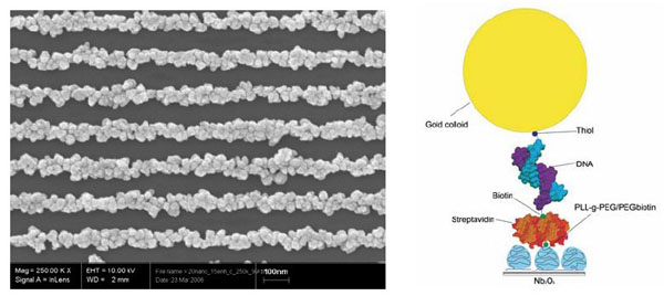
The combination of these novel top-down and bottom-up approaches enables us to interact with complex biological systems: tissues, cells, proteins and DNA in an unprecedented manner. In addition, new tools, such as arrays of nanoparticles and nanowires can be created on a large scale with promising applications in electronic and optical biosensing. (See figure below).
Examples for the handling of natural and artificial macromolecular complexes at the nanometer scale will be presented. Proteins, vesicles, macromolecular assemblies, and nanoparticles specifically placed onto predefined artificial patterns can trigger defined functions in cells, reveal the details of cell-surface interactions and allow for the ultimate miniaturization of array-type sensors down to the single molecule level. Recently, we have put a lot of efforts into achieving not only spatial but also a dynamic control over the properties of biointerfaces. Surfaces that change upon external stimuli provide us with new research tools for studying complex biological problems and for tissue engineering. Highlights for the use of novel, electronically- or photo-active surfaces for applications in biosensing and local drug delivery will also be presented.
About the speaker: János Vörös is an Associate Professor in the Institute for Biomedical Engineering of the University and ETH Zurich (Department for Information Technology and Electrical Engineering) heading the Laboratory for Biosensors and Bioelectronics since 1st of January, 2006.
Born on June 24th of 1970 in Keszthely, Hungary, János Vörös has studied Physics at the Eötvös Loránd University in Budapest. After receiving a diploma in Physics in 1995, he was a doctoral student at the Department of Biological Physics of the Eötvös University (in collaboration with Microvacuum Ltd.) where he received his PhD in Biophysics in 2000. Since 1998 he was a member of the BioInterface group in the Laboratory for Surface Science and Technology at the Department of Materials of ETH Zurich as visiting scientist, postdoc, and from 2004 as group leader of the Dynamic BioInterfaces group until 2006. Prof. Vörös also has an adjunct appointment at the Department of Engineering Science and Mechanics of the Pennsylvania State University.
Prof. Vörös is interested in research and teaching in the areas of Bioelectronics, Nano-Biotechnology, Biosensors, Biophysics, and Biomaterials with special focus on the understanding, monitoring and controlling of molecular and cellular processes at biological interfaces. His research group focuses on the development of novel microarray based biosensor techniques for diagnostics and drug discovery in collaboration with academic and industrial partners, as well as on using nanobiotechnology for interfacing neural networks and controlling synapse formation.
Download visuals (14.1 MB pdf)
Secondary navigation
- January 29, 2018
- August 30, 2017
- Past seminars
- 2016 - 2017 Seminars
- 2015 - 2016 Seminars
- 2014 - 2015 Seminars
- 2013 - 2014 Seminars
- 2012 - 2013 Seminars
- 2011 - 2012 Seminars
- 2010 - 2011 Seminars
- 2009 - 2010 Seminars
- 2008 - 2009 Seminars
- 2007 - 2008 Seminars
- 2006 - 2007 Seminars
- August 31, 2007
- June 29, 2007
- June 20, 2007
- June 5, 2007
- May 30, 2007
- May 16, 2007
- May 15, 2007
- April 24, 2007
- March 27, 2007
- March 14, 2007
- February 9, 2007
- February 8, 2007
- January 12, 2007
- December 5, 2006
- November 14, 2006
- October 31, 2006
- October 27, 2006
- October 26, 2006
- October 20, 2006
- September 20, 2006
- September 20, 2006
- September 20, 2006
- September 19, 2006
- 2005 - 2006 Seminars
- August 23, 2006
- August 22, 2006
- June 26, 2006
- June 20, 2006
- June 16, 2006
- June 7, 2006
- June 6, 2006
- May 30, 2006
- May 17, 2006
- May 10, 2006
- April 27, 2006
- April 12, 2006
- March 31, 2006
- March 29, 2006
- March 22, 2006
- March 15, 2006
- February 27, 2006
- February 8, 2006
- January 25, 2006
- January 19, 2006
- January 18, 2006
- January 17, 2006
- January 11, 2006
- November 30, 2005
- November 23, 2005
- November 2, 2005
- October 26, 2005
- October 25, 2005
- October 5, 2005
- September 28, 2005
- 2005 Seminars
Links